The UNC School of Medicine laboratory of Camille Ehre, produced striking images of respiratory tract produced by infected respiratory epithelial cells. Ehre, captured these images to illustrate how intense the infection of the airways can be in very graphic and easily understood images.
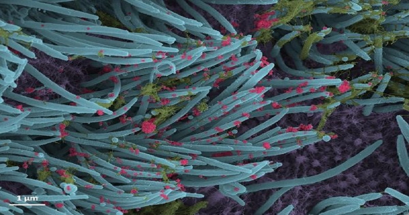
These cells were examined 96 hours later, using scanning electron microscopy. The images, re-colourized, show infected ciliated cells with strands of mucus attached to the cilia tips. Cilia are hair-like structures on the surface of airway epithelial cells that transport mucus from the lungs. A higher power magnification image shows the structure and density of SARS-CoV-2 virions produced by human airway epithelia. Virions are the complete, infectious form of the virus released onto respiratory surfaces by infected host cells.
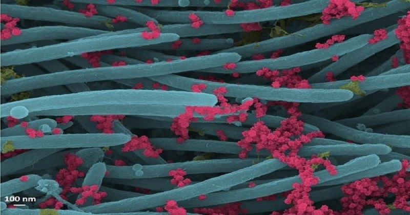
These images can help researchers understand the viral load or burden of SARS-CoV-2, which can also help determine how likely a person is to develop severe COVID-19 and transmit it to others. These images also support the studies that lay emphasis on the need to wear masks to slow down the transmission of the virus.
Post Your Comments